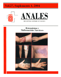
Classification of vascular anomalies (tumours and malformations). Clinical characteristics and natural history
Keywords:
Hemangioma. Malformación vascular. Clasificación.Abstract
Vascular anomalies are divided into tumours and malformations. Haemangiomas are the most frequent amongst the former. Not normally present at birth, except in a premonitory form, they grow for 10-12 months due to hyperplasia, to subsequently undergo a progressive involution for a period that might last from ten to twelve years. They have an incidence of up to 12% in newborns; they are more common amongst girls; and are divided into superficial, deep and compound. Congenital haemangiomas and those that do not undergo involution are considered to be rare entities. Vascular malformations, with a lower incidence than haemangiomas, are always present at birth, they grow by hypertrophy and never undergo involution. According to the classification of the ISSVA, vascular malformations are divided - depending on the vessel affected - into capillary or venular (port-wine stain), venous, lymphatic, arteriovenous and combined or complex. Each of these has certain defining clinical and haemodynamic peculiarities. Within the final group are included some with a low flow, such as the Klippel-Trenaunay syndrome (venous and lymphatic venular vascular malformation associated with the muscular-skeletal hypertrophy of an extremity), and others with a high flow, such as the Parkes-Weber syndrome.Downloads
Download data is not yet available.
Downloads
Published
2008-12-22
How to Cite
1.
Redondo P. Classification of vascular anomalies (tumours and malformations). Clinical characteristics and natural history. An Sist Sanit Navar [Internet]. 2008 Dec. 22 [cited 2026 Feb. 21];27:9-25. Available from: https://recyt.fecyt.es/index.php/ASSN/article/view/5032
Issue
Section
Research articles
License
La revista Anales del Sistema Sanitario de Navarra es publicada por el Departamento de Salud del Gobierno de Navarra (España), quien conserva los derechos patrimoniales (copyright ) sobre el artículo publicado y favorece y permite la difusión del mismo bajo licencia Creative Commons Reconocimiento-CompartirIgual 4.0 Internacional (CC BY-SA 4.0). Esta licencia permite copiar, usar, difundir, transmitir y exponer públicamente el artículo, siempre que siempre que se cite la autoría y la publicación inicial en Anales del Sistema Sanitario de Navarra, y se distinga la existencia de esta licencia de uso.