Imaging studies in the diagnosis of haemangiomas and vascular malformations
Keywords:
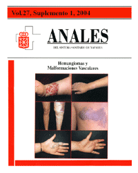
Hemangioma. Malformación vascular. Diagnóstico.Abstract
The diagnosis of haemangiomata and cutaneous vascular malformations is based on clinical history and physical exploration. Imaging studies might be necessary to clarify and confirm the diagnosis, and in order to analyse the extent of the lesions by permitting an evaluation of their non-visible component as well as the affection of neighbouring structures. Finally, they also play an important role when it comes to planning and directing treatment, whether surgical or endovascular. The imaging techniques employed for haemangiomas and vascular malformations include: plain films ultrasound (Doppler), computerised tomography (Angio-CT), magnetic resonance (Angio-MR) and the angiographic techniques (arteriography, phlebography).Downloads
Download data is not yet available.
Downloads
Published
2008-12-22
How to Cite
1.
Martínez de la Cuesta A. Imaging studies in the diagnosis of haemangiomas and vascular malformations. An Sist Sanit Navar [Internet]. 2008 Dec. 22 [cited 2026 Feb. 1];27:71-80. Available from: https://recyt.fecyt.es/index.php/ASSN/article/view/5037
Issue
Section
Research articles
License
La revista Anales del Sistema Sanitario de Navarra es publicada por el Departamento de Salud del Gobierno de Navarra (España), quien conserva los derechos patrimoniales (copyright ) sobre el artículo publicado y favorece y permite la difusión del mismo bajo licencia Creative Commons Reconocimiento-CompartirIgual 4.0 Internacional (CC BY-SA 4.0). Esta licencia permite copiar, usar, difundir, transmitir y exponer públicamente el artículo, siempre que siempre que se cite la autoría y la publicación inicial en Anales del Sistema Sanitario de Navarra, y se distinga la existencia de esta licencia de uso.